Visualizzazione post con etichetta Oncology. Mostra tutti i post

lunedì 9 luglio 2012

What is metastatic cancer (Definition)


Metastatic cancer is a cancer that has spread from the part of the body where it started (the primary site) to other parts of the body. When cancer cells break away from a tumor, they can travel to other areas of the body through either the bloodstream or the lymph system (a collection of vessels that carry fluid and immune system cells).
Image of the lymph system.
    The Lymph System
If the cells travel through the lymph system, they may end up in the lymph nodes (small, bean-sized collections of immune cells) or spread to other organs. If the cells travel through the bloodstream they can go to any part of the body. Most often, the cancer cells break off and travel in the bloodstream. Many of these cells die, but some settle in a new area, begin to grow, and form new tumors. This spread of cancer to a new part of the body is called metastasis.
In order for cancer cells to spread to new parts of the body, they have to go through several changes. They have to be able to break away from the original tumor and enter the bloodstream or lymph system, which can carry them to another part of the body. At some point they need to attach to the wall of a blood or lymph vessel and move through it into a new organ. They then need to be able to grow and thrive in their new location. All the while, they need to be able to avoid attacks from the body's immune system. Going through all these steps means the cells that start new tumors may no longer be exactly the same as the ones in the tumor they started in. This may make treatment more difficult.
Even when cancer has spread to a new area, it is still named after the part of the body where it started. For example, if prostate cancer spreads to the bones, it is still called prostate cancer (not bone cancer). Likewise, breast cancer that has spread to the lungs it is still called breast cancer, not lung cancer.
Sometimes the metastatic tumors have already begun to grow when the cancer is first found and diagnosed. And in some cases, a metastasis may be found before the original (primary) tumor is found. If a cancer has already spread to many places before it is found, it may be hard to figure out where it started. If this happens the cancer is called cancer of unknown primary.

lunedì 5 dicembre 2011

Is A Mole Benign Or Cancerous? New Staining Test Can Provide New Measure

Up till now, finding out if a mole is a potentially life-threatening melanoma has been a complex task, resulting in both under- and over diagnosis. However, a novel diagnostic staining test has been designed, developed and studied by investigators at Weill Medical College. According to the researchers, the test can provide a new measure of determining whether a mole is benign or cancerous, and has the potential for expanded use across all cancers. The study is published in the Nov. 21 issue of the Archives of Dermatology.

The researchers explain that the novel test, which relies on the soluble adenylyl cyclase (sAC) expression pattern, provides objective results - melanoma is present if sAC is present in the nucleus of cells from a skin biopsy, and benign if the nucleus is not positive.

The majority of diagnostic stains currently used highlight a particular cell in the biopsy. If the stain is more intense it tends to be melanoma, but the definition of 'intense' is subjective.


There are various types of moles, this one is a intradermal nevus

Senior author Dr. Jonathan Zippin, explains:

"The sAC stain is either positive or negative in the cell's nucleus. Other stains require an interpretation of staining intensity, which means that a diagnosis of melanoma can rest on a pathologist's opinion.

As a clinical dermatologist, this uncertainty is difficult to deal with. No one wants to miss a true diagnosis of melanoma, but telling someone they have melanoma, when they may not, changes his or her life."


In order to diagnose melanoma, the skin lesion, together with an area of healthy tissue around it, must be surgically removed, which can result in disfiguration. According to Dr. Zippin, those who are diagnosed with melanoma are concerned they might develop other forms of cancer, as individuals' diagnoses with the disease have a greater risk of developing another cancer.

sAC stain should be used together with other diagnostic tests, such as light microscopy and three other strains currently available, according to the investigation's lead author, Dr. Cynthia Margo.

Dr. Zippin, said:

"What I hope is that five years down the line, this and other stains will help pathologists remove any uncertainty as to whether a biospy is worrisome."


Dr. Margo explains, however, that the sAC provides one benefit that other stains do not. "The greatest practical application of sAC is using it to determine the cancer-free margins of a lentigo maligna that needs to be removed. This is a common melanoma in situ, usually found on the face, that does not show invasive growth. But until the development of sAC, it was very difficult to tell where the cancerous lesion ended." The sAC stain, which Margo uses on several patients, "is vastly superior to any other test we have now to determine margins."

Dr. Zippin explains:

"The new stain is a monoclonal antibody that binds to sAC, which is a signaling molecule," who has studied the molecule. He continues: "sAC senses changes in the interior of cells, such as pH and metabolism, and responds by regulating gene expression, metabolism and growth."


The sAC protein was discovered by Dr. Jochen Buck and Dr. Lonny Levin, both professors of pharmacology at Weill Cornell Medical College.

Dr. Zippin and Dr. Margo developed the antibody used to develop the diagnostic test. As their research gained potential, Dr. Zippin, Dr. Margo, Dr. Buck and Dr. Levin created a company called Cutting Edge Pattern Biotech in order to share the antibody with the scientific community. At present, they are scientific advisers but have no fiscal role in the company.

Dr. Zippin said:

"Beause the sAC protein is expressed in all tissues of the body, and appears to be linked to processes important for the development of cancer, we predict that immunostain will be useful for the diagnosis of many other cancers and diseases."


Even though the diagnostic stain does not require approval from the FDA, the investigators explain that Cutting Edge Pattern Biotech plans to seek federal approval of the sAC antibody as a diagnostic test.

Key To Melanoma Metastasis

Researchers from UNC Lineberger Comprehensive Cancer Center are part of a team that has identified a protein, called P-Rex1, that is key to the movement of cells called melanoblasts. When these cells experience uncontrolled growth, melanoma develops.

Melanoma is one of the only forms of cancer that is still on the rise and is one of the most common forms of cancer in young adults. The incidence of melanoma in women under age 30 has increased more than 50 percent since 1980. Metastases are the major cause of death from melanoma.

The team found that mice lacking the P-Rex1 protein are resistant to melanoma metastases. When researchers tested human melanoma cells and tumor tissue for the protein, P-Rex1 was elevated in the majority of cases - a clue that the protein plays an important role in the cancer's spread. Their findings were published in the journal Nature Communications.

"We know that mutations in a gene called BRAF are important for the development of melanoma and several years ago we published a collaborative paper listing 82 proteins that seem to be affected by this genetic pathway. From that list, we focused on P-Rex1 in collaboration with Dr. Nancy Thomas here at UNC and researchers in the United Kingdom," says Channing Der, PhD, a member of the UNC research team. Der is Kenan Professor of pharmacology at UNC-Chapel Hill and member of UNC Lineberger.

A drug approved this summer, vemurafenib, is the first treatment directed at the BRAF mutation. Clinical trials found that the treatment offers a significant survival benefit.

"We think that vemurafenib may work, in part, by blocking the up-regulation of P-Rex1," Der adds.

"As a physician and scientist, I know firsthand the frustration of having very limited therapeutic options to offer to patients with metastatic melanoma," says Nancy Thomas, MD, PhD, whose laboratory analyzed the protein's expression in human cells. "Pinpointing that P-Rex1 plays a key role in metastasis gives us a better understanding of how vemurafenib may work and a target for developing new treatments," she adds.

Metastatic Breast And Ovarian Cancer Vaccine - Promising Results

A trial published in Clinical Cancer Research demonstrated a positive response in both metastatic breast cancer and ovarian cancer to a recombinant poxviral vaccine.

Lead investigator James Gulley, M.D., Ph.D., director and deputy chief of the clinical trials group at the Laboratory of Tumor Immunology and Biology at the National Cancer Institute commented:

"With this vaccine, we can clearly generate immune responses that lead to clinical responses in some patients."


For the trial, funded by the National Cancer Institute, Gulley and his team involved 26 patients, all of them heavily pretreated, with 21 patients receiving at least three prior chemotherapy treatments, and designated them to receive monthly vaccinations with PANVAC vaccine that contains transgenes for MUC-1, CEA and three T cell costimulatory molecules.

In 12 breast cancer patients the team established an average time to disease progression of 2.5 months and an average overall survival rate of 13.7 months. In four patients the disease was stable. The average time to progression for the 14 ovarian cancer patients was two months and the average survival rate of 15 months.

The most common side effect after treatment was mild reactions at the injection-site.

Because of the rising interest in cancer vaccinations, Gulley recommends for further studies to be conducted in order to establish which vaccines are beneficial for particular patients.

He concludes:

"The sustained benefit seen in some patients in this study underscores the potential for therapeutic vaccines to impact clinical outcomes without toxicity. However, more studies in the appropriate patient populations are required to adequately assess efficacy."

Wi-Fi Laptops Harm Sperm Motility And Increase Sperm DNA Fragmentation

Males who place a laptop on their laps with the WI-FI on might have a greater risk of reduced sperm motility and more sperm DNA fragmentation, which could, in theory, undermine their chances of becoming fathers, researchers from Nascentis Medicina Reproductiva, Argentina, and the Eastern Virginia Medical School, USA, reported in the journal Fertility and Sterility this week. Sperm motility refers to the percentage of sperm in a semen sample that are moving - normally, a high percentage of all sperm should be moving (thrashing their tails and swimming).

This study was done in an artificial setting. The male participants were not tested with the laptops on their laps - semen samples were taken, placed under laptops for four hours, and then analyzed.

Previous studies had already shown that placing a laptop on a man's lap could potentially affect his fertility, especially if this occurs frequently and for long periods. The laptop can cause scrotal hyperthermia (elevated testicle temperature), which can considerably affect the quality of his sperm (Link to 2010 study).

In this new study, the authors explain that not only might the laptop-on-lap undermine semen quality, but also the Wi-Fi, if the laptop is near semen. They found that there was less damage when there was no Wi-Fi signal than when there was.

The double-whammy of the Wi-Fi signal and laptop temperature can cause:
A decrease in human sperm motility
Sperm DNA fragmentation - irreversible changes in the genetic code
Perhaps the electromagnetic radiation emitted by Wi-Fi damages the semen, the scientists suggested.

Wi-Fi stands for "wireless fidelity". The term refers to a group of technical standards which enable the transmission of data over wireless networks. Put simply: Wi-Fi means wireless internet connection.

Conrado Avendaño and team carried out a study involving semen samples from 29 healthy and fertile males. They experimented on their semen samples in two environments:
The Wi-Fi sample. A few drops of semen were place under a laptop with the Wi-Fi switched on. The laptop was downloading data from the internet non-stop.
The non-Wi-Fi sample. Identical to the environment above, but with no Wi-Fi switched on.
Four hours later, they found:
One quarter of the sperm had lost motility in the Wi-Fi samples
14% of the sperm had lost motility in the non-Wi-Fi samples
9% of the sperm showed DNA damage in the Wi-Fi samples
3% of the sperm showed DNA damage in the non-Wi-Fi sampes
Avendaño said:

"Our data suggest that the use of a laptop computer wirelessly connected to the internet and positioned near the male reproductive organs may decrease human sperm quality.
,br> At present we do not know whether this effect is induced by all laptop computers connected by Wi-Fi to the internet or what use conditions heighten this effect."


The authors carried out a separate test to determine what the EM radiation levels might be near a Wi-Fi connected laptop and a non-Wi-Fi one. The difference was significant - when the computer was not Wi-Fi connected EM radiation readings were "negligible".

According to PC Mag, Electromagnetic Radiation, also known as EM Radiation, or EMR is:

"The energy that radiates from all things in nature and from man-made electronic systems. It includes cosmic rays, gamma rays, x-rays, ultraviolet light, visible light, infrared light, radar, microwaves, TV, radio, cellphones and all electronic transmission systems. Electromagnetic radiation is made up of electric and magnetic fields that move at right angles to each other at the speed of light."


The authors, as well as other experts who have commented on this study, stress that until a long-term study with a larger group of men and in natural environments is carried out, this one is only "interesting" and not really biologically relevant for humans.

Even though previous studies have looked at laptop usage and sperm quality, none have determined whether there is any impact on how many children men subsequently have or don't have.

In an Abstract in the journal, the authors concluded:

"To our knowledge, this is the first study to evaluate the direct impact of laptop use on human spermatozoa. Ex vivo exposure of human spermatozoa to a wireless internet-connected laptop decreased motility and induced DNA fragmentation by a nonthermal effect.

We speculate that keeping a laptop connected wirelessly to the internet on the lap near the testes may result in decreased male fertility. Further in vitro and in vivo studies are needed to prove this contention."

Coffee Emerges As Protective Against Cancer And Other Diseases

Starbucks fans around the world can rejoice that their tipple gets the thumbs up yet again. Already shown to protect against a number of diseases, a recent study in the Cancer Epidemiology, Biomarkers & Prevention, a journal of the American Association for Cancer Research, shows coffee drinkers who consume more than four cups a day have a 25% lower risk of developing Endometrial Cancer. It is thought that the antioxidant properties in coffee may be a part of the mechanism.

Edward Giovannucci, M.D., Sc.D., professor of nutrition and epidemiology at the Harvard School of Public Health, and a senior researcher on the study, said coffee is starting to be proven as a protective agent in cancers that are linked to obesity, estrogen and insulin.

He said :

"Coffee has already been shown to be protective against diabetes due to its effect on insulin ... So we hypothesized that we'd see a reduction in some cancers as well."


Giovannucci, and his colleagues, including Youjin Je, a doctoral candidate in his lab, looked at endometrial cancer cases in nearly 70,000 women who enrolled in the Nurse's Health Study. Over 26 years they documented 672 cases of endometrial cancer. More than four cups of coffee was linked with a 25% decrease in risk, while two to three cups had a 7% lower risk.

The results seemed to hold true for decaf as well, with a 22% reduction for more than two cups per day.

Giovannucci said he'd like to see further research on the effects of coffee on cancer, because in this and similar studies, coffee intake is self-selected and not randomized.

He said :

"Coffee has long been linked with smoking, and if you drink coffee and smoke, the positive effects of coffee are going to be more than outweighed by the negative effects of smoking ... However, laboratory testing has found that coffee has much more antioxidants than most vegetables and fruits."

domenica 27 novembre 2011

Benign Prostatic Hypertrophy Treatment & Management

Approach Considerations

Patients with mild symptoms (IPSS/AUA-SI score < 7) or moderate-to-severe symptoms (IPSS/AUA-SI score ≥8) of benign prostatic hyperplasia (BPH) who are not bothered by their symptoms and are not experiencing complications of BPH should be managed with a strategy of watchful waiting. In these situations, medical therapy is not likely to improve their symptoms and/or quality of life (QOL). In addition, the risks of treatment may outweigh any benefits. Patients managed expectantly with watchful waiting are usually re-examined annually.
Transurethral resection of the prostate (TURP) has long been accepted as the criterion standard for relieving bladder outlet obstruction (BOO) secondary to BPH. In current clinical practice, most patients with BPH do not present with obvious surgical indications; instead, they often have milder lower urinary tract symptoms (LUTS) and, therefore, are initially treated with medical therapy.
The era of medical therapy for BPH dawned in the mid 1970s with the use of nonselective alpha-blockers such as phenoxybenzamine. The medical therapeutic options for BPH have evolved significantly over the last 3 decades, giving rise to the receptor-specific alpha-blockers that comprise the first line of therapy.

Alpha-1–Receptor Blockade in Benign Prostatic Hyperplasia

A significant component of LUTS secondary to BPH is believed to be related to the smooth-muscle tension in the prostate stroma, urethra, and bladder neck. The smooth-muscle tension is mediated by the alpha-1-adrenergic receptors; therefore, alpha-adrenergic receptor–blocking agents should theoretically decrease resistance along the bladder neck, prostate, and urethra by relaxing the smooth muscle and allowing passage of urine.
BPH is predominantly a stromal proliferative process, and a significant component of prostatic enlargement results from smooth-muscle proliferation. The stromal-to-epithelial ratio is significantly greater in men with symptomatic BPH than in those with asymptomatic BPH.
The 3 subtypes of the alpha-1 receptor include 1a, 1b, and 1c. Of these, the alpha-1a receptor is most specifically concentrated in the bladder neck and prostate. Provided that the alpha-1a subtype is predominant in the prostate, bladder neck, and urethra, but not in other tissues, drugs that are selective for this receptor (ie, tamsulosin) may have a potential therapeutic advantage.
Tamsulosin is considered the most pharmacologically uroselective of the commercially available agents because of its highest relative affinity for the alpha-1a receptor subtype. In 2008, the US Food and Drug Administration (FDA) approved a new alpha-1a receptor selective blocker, silodosin (Rapaflo). It is indicated for treatment of the signs and symptoms of BPH.
The efficacy of the titratable alpha-blockers doxazosin and terazosin (Hytrin) is dose-dependent. Maximum tolerable doses have not been defined for any alpha-blocker; however, the higher the dose, the more likely the adverse events (orthostatic hypotension, dizziness, fatigue, ejaculatory disorder, nasal congestion). Despite the requirement for dose titration and blood pressure monitoring, these older, often less costly, alpha-blockers appear to be equally effective to tamsulosin and alfuzosin, and the 2010 AUA guidelines state that they remain reasonable choices for patients with moderate-to-severe LUTS due to BPH.[2]
An approximately 4- to 6-point improvement is expected in IPSS/AUA-SI scores when alpha-blockers are used. Interestingly, alpha-blocker therapy has not been shown to reduce the overall long-term risk for acute urinary retention (AUR) or BPH-related surgery.[7]
Hellstrom and Sikka reported in 2006 that the acute administration of tamsulosin effects ejaculatory function and ejaculate volume. Nearly 90% of study subjects experienced decreased ejaculate volume, and approximately 35% experienced anejaculation. In their study, subjects treated with alfuzosin or placebo did not experience anejaculation.[8]

Alpha-adrenergic receptor blockers

The alpha-blocking agents administered in BPH studies can be subgrouped according to receptor subtype selectivity and the duration of serum elimination half-lives, as follows:
  • Nonselective alpha-blockers - phenoxybenzamine
  • Selective short-acting alpha-1 blockers - prazosin, alfuzosin, indoramin
  • Selective long-acting alpha-1 blockers - terazosin, doxazosin, slow-release (SR) alfuzosin.
  • Partially subtype (alpha-1a)–selective agents – tamsulosin, silodosin

Nonselective alpha-blockers

Phenoxybenzamine was the first alpha-blocker studied for BPH. It is nonselective, antagonizing both the alpha 1- and alpha 2-adrenergic receptors, which results in a higher incidence of adverse effects. Because of the availability of more alpha-1-receptor–specific agents, phenoxybenzamine is currently not often used for the treatment of BPH. The 2010 update to the AUA guideline for BPH retains the statement that insufficient data exist for a recommendation of phenoxybenzamine or of prazosin for treatment of LUTS secondary to BPH. This statement was originally published in the 2003 AUA BPH guidelines.[2]

Phosphodiesterase-5 enzyme inhibitors

Statistically significant symptomatic improvements have been reported for patients with BPH receiving tadalafil. It has also been approved for the treatment of simultaneous BPH and erectile dysfunction (ED). Phosphdiesterase-5 (PDE5) inhibitors are known to mediate smooth muscle relaxation in the lower urinary tract.

Intraoperative floppy iris syndrome

Intraoperative floppy iris syndrome (IFIS) is characterized by miosis, iris billowing, and prolapse in patients undergoing cataract surgery who have taken or currently take alpha-1-blockers. It is particularly prevalent among patients taking tamsulosin. The 2010 AUA guideline recommends that clinicians ask patients about planned cataract surgery when offering alpha-blocker therapy for LUTS due to BPH. Alpha-blockers should not be initiated until cataract surgery is completed.[2] Patients currently on alpha-blocker therapy must disclose this to their ophthalmologist prior to cataract surgery so that the appropriate preoperative and intraoperative precautions can be taken. Experienced ophthalmologists can thereby reduce the risk of complications from IFIS.[9, 2]
In a review by Bell et al, exposure to tamsulosin within 14 days of cataract surgery was significantly associated with serious postoperative ophthalmic adverse events, specifically IFIS and its complications (ie, retinal detachment, lost lens or fragments, endophthalmitis). No significant associations were noted with exposure to other alpha-blocker medications or to previous exposure to tamsulosin or other alpha-blockers.[10]

5-Alpha-Reductase Inhibitors in Benign Prostatic Hyperplasia

Hormonal medical management emerged from the discovery of a congenital form of pseudohermaphroditism secondary to DHT deficiency (due to a lack of 5-alpha-reductase activity). This deficiency produced a hypoplastic prostate. The two types of 5-alpha-reductase include type 1 (predominantly located in extraprostatic tissues, such as skin and liver) and type 2 (predominant prostatic reductase).
Inhibition of 5-alpha-reductase type 2 blocks the conversion of testosterone to DHT, resulting in lower intraprostatic levels of DHT. This leads to inhibition of prostatic growth, apoptosis, and involution. The exact role of 5-alpha-reductase type 1 in normal and abnormal prostatic development is undefined. 5-Alpha-reductase inhibitors improve LUTS by decreasing prostate volumes; thus, patients with larger prostates may achieve a greater benefit. Further, maximal reduction in prostate volume requires 6 months of therapy.

5-Alpha reductase inhibitors

Finasteride (Proscar), a 4-aza-steroid, has demonstrated 5-alpha type II–blocking activity, resulting in the inhibition of DHT-receptor complex formation. This effect causes a profound decrease in the concentration of DHT intraprostatically, resulting in a consistent decrease in prostate size. One third of men treated with this agent exhibit improvements in urine flow and symptoms.
Dutasteride (Avodart) has an affinity for both type 1 and type 2 5-alpha-reductase receptors. The significance of blockage of type 1 receptors is currently unknown.
Both finasteride and dutasteride actively reduce DHT levels by more than 80%, improve symptoms, reduce the incidence of urinary retention, and decrease the likelihood of surgery for BPH. Adverse effects are primarily sexual in nature (decreased libido, erectile dysfunction, ejaculation disorder).
Both finasteride and dutasteride may reduce serum prostate-specific antigen (PSA) values by as much as 50%. The decrease in PSA is typically maximally achieved when the maximal decrease in prostatic volume is noted (6 months). Thus, one must take this into account when using PSA to screen for prostate cancer.
One prospective, randomized, double-blind study by the Enlarged Prostate International Comparator Study (EPICS) was conducted to compare the efficacy of dutasteride to that of finasteride in men with symptomatic BPH. While this study was conducted over the course of only one year, the data suggest that both of these drugs were similarly effective in reducing prostate volume, improving Qmax, and LUTS for this population. The long-term outcomes are yet to be investigated.[11]
Because these drugs interfere with the metabolism of testosterone, they are contraindicated in children and pregnant females. In addition, pregnant females or those who are considering conception should not handle crushed or broken tablets because of the potential for absorption and subsequent potential risk to a male fetus.
In patients with LUTS and enlarged prostates, 5-alpha-reductase inhibitors are believed to be appropriate and effective treatment.

5-Alpha reductase inhibitors and prostate cancer

On June 9, 2011, the US Food and Drug Administration announced revisions to the prescribing information for 5-alpha reductase inhibitors (5-ARIs). These agents include finasteride (Proscar, Propecia) and dutasteride (Avodart, Jalyn). 5-ARIs are indicated for benign prostatic hypertrophy and alopecia.
Data from 2 large, randomized, controlled trials observed an increased risk of being diagnosed with a more serious form of prostate cancer (high-grade prostate cancer) in trial participants taking 5-ARIs. The 2 trials are the Prostate Cancer Prevention Trial (PCPT) and the Reduction by Dutasteride of Prostate Cancer Events (REDUCE) trial. Both of these trials observed a decreased incidence of prostate cancer overall when preventive treatment included 5-ARIs, but an increased incidence of high-grade prostate cancer in men taking dutasteride or finasteride compared with placebo.[12, 13]
The revised prescribing information recommends that prior to initiating therapy with 5-ARIs, perform appropriate evaluations to rule out other urological conditions, including prostate cancer, that might mimic benign prostatic hyperplasia (BPH).

Combination Therapy

The alpha-1-receptor blockers provide rapid relief, while the 5-alpha-reductase inhibitors target the underlying disease process.[7] The Medical Therapy of Prostatic Symptoms (MTOPS) trial demonstrated that combination therapy reduced the risk of progression and produced a greater improvement in IPSS than therapy with finasteride or doxazosin alone. The risks of AUR and BPH-related surgery were reduced with combination therapy or finasteride in comparison with doxazosin monotherapy.[14]
The Symptom Management After Reducing Therapy (SMART-1) trial demonstrated that after 6 months of combination therapy, discontinuation of the alpha-1-blocker is possible in men with moderate LUTS. However, those with severe LUTS may require longer combination therapy.[14]

Anticholinergic Agents

Historically, anticholinergics were discouraged in men with BPH because of concerns of inducing urinary retention. Trials have demonstrated a slight increase in PVR; however, AUR rates were low. Importantly, these trials consisted of patients with low baseline PVR.
The 2010 AUA BPH guidelines recommend anticholinergic agents for management of LUTS in patients who do not have an elevated PVR and whose LUTS are primarily irritative. Baseline PVR should be obtained prior to initiation of anticholinergic therapy, to assess for urinary retention.[15] Caution with anticholinergics is recommended with patients whose PVR is greater than 250-300 mL.[2]

Landmark Clinical Trials

Numerous phase II and phase III trials of drugs used in the treatment of BPH have been conducted. A few landmark studies are selected below.
The Proscar Long-Term Efficacy and Safety Study (PLESS), patients treated with finasteride (5 mg/d) were at a significantly lower risk of developing AUR or needing surgery.[16] This was a multicenter, 4-year, double-blind, placebo-controlled study of 3,040 men. Men with PSA levels of more than 10 ng/mL and those with prostate cancer were excluded.
The Medical Therapy of Prostatic Symptoms (MTOPS) trial demonstrated that combination therapy with doxazosin and finasteride was well tolerated, and was superior to placebo and monotherapy with either agent. The primary endpoints of the study were reduction in AUA-SI score, AUR, recurrent infections, renal insufficiency, incontinence, changes in flow, and PSA level and a lower rate of invasive treatments. MTOPS was a multicenter, 4- to 6-year, double-blind, randomized, placebo-controlled trial of 3,047 men with symptomatic BPH.[17]
In the Alfuzosin Long-Term Efficacy and Safety Study (ALTESS), alfuzosin (10 mg/d) decreased the risk of LUTS deterioration and significantly improved QOL and peak urinary flow rate. ALTESS was a 2-year, double-blind, placebo-controlled study of 1,522 men. Notably, these men had greater risk factors for BPH progression (ie, older age, higher IPSS scores, larger prostate size, lower Qmax, and higher PVR) than those in the MTOPS trial. Alfuzosin did not reduce the risk of AUR but tended to reduce the risk of surgery.[18]
In the international real-life practice study of alfuzosin once daily (ALF-ONE), 3 years of alfuzosin (10 mg/d) decreased IPSS by one third, with significant improvements in nocturia and bother score. ALF-ONE was conducted in 689 European men with a mean age of 67.6 years. Clinical progression of worsening of IPSS (≥4 points) was seen in 12.4%, AUR in 2.6%, and requirement of BPH-related surgery in 5.7%. Alfuzosin was well tolerated, with dizziness the most common adverse effect (4.5%). Notably, symptom worsening during treatment and high PSA levels appeared to be the best predictors of clinical progression.[19]
Four-year results in the Combination of Avodart and Tamsulosin (CombAT) study revealed that for men with prostate volumes of 30-58 mL, combination therapy with dutasteride (dual 5-alpha-reductase inhibitor) and tamsulosin (alpha-1-blocker) improved symptoms, urinary flow, and QOL better than monotherapy with either drug, although not in men who had a prostate volume of 58 mL or more.[20] The adverse-effect profile of combination therapy was similar to that of monotherapy, although drug-related adverse events were more common with combination therapy.[21] CombAT is a 4-year, multicenter, randomized, double-blind, parallel group study of 4,844 men aged 50 years or older with moderate-to-severe BPH symptoms (IPSS ≥12), prostate volume of 30 mL or greater, and a PSA level of 1.5-10 ng/mL. This study contributes to the standard of care shifting towards combined drug therapy in appropriately selected patients, while better defining the role of the alpha-blockers.[15]

Phytotherapeutic Agents and Dietary Supplements

Phytotherapeutic agents and dietary supplements are considered emerging therapy by the AUA Guidelines panel and are not recommended for the treatment of BPH because of the lack of evidence at this time.
Pharmaceuticals derived from plant extracts are widely used throughout the world for the treatment of various medical ailments. In 1998, Americans spent a total of $3.65 billion on all herbal remedies. In France and Germany, plant extracts have a market share of up to 50% of all drugs prescribed for symptomatic BPH. In the United States, these agents are also popular and readily available.
The attraction to phytotherapeutic agents appears to be related to the perception of therapeutic healing powers of natural herbs, the ready availability, and the lack of adverse effects.
Most of the phytotherapeutic agents used in the treatment of LUTS secondary to BPH are extracted from the roots, seeds, bark, or fruits of plants listed below. Some suggested active components include phytosterols, fatty acids, lectins, flavonoids, plant oils, and polysaccharides. Some preparations derive from a single plant; others contain extracts from 2 or more sources.
Each agent has one or more proposed modes of action. The following modes of action are suggested:
  • Antiandrogenic effect
  • Antiestrogenic effect
  • Inhibition of 5-alpha-reductase
  • Blockage of alpha receptors
  • Antiedematous effect
  • Anti-inflammatory effect
  • Inhibition of prostatic cell proliferation
  • Interference with prostaglandin metabolism
  • Protection and strengthening of detrusor
The origins of phytotherapeutic agents are as follows:
  • Saw palmetto, (American dwarf palm; Serenoa repens, Sabal serrulata) fruit
  • South African star grass (Hypoxis rooperi) roots
  • African plum tree (Pygeum africanum) bark
  • Stinging nettle (Urtica dioica) roots
  • Rye (Secale cereale) pollen
  • Pumpkin (Cucurbita pepo) seeds

Saw palmetto (American dwarf palm)

Extracts of saw palmetto berries are the most popular botanical products for BPH. The active components are believed to be a mixture of fatty acids, phytosterols, and alcohols. The proposed mechanisms of action are antiandrogenic effects, 5-alpha-reductase inhibition, and anti-inflammatory effects.
The recommended dosage is 160 mg orally twice daily. Studies show significant subjective improvement in symptoms without objective improvements in urodynamic parameters. Minimal adverse effects include occasional GI discomfort.
The 2010 AUA guidelines, based on more recent studies, do not detect a clinically meaningful effect of saw palmetto on LUTS. Further clinical trials are underway.[2] In fact, in a double-blind, multicenter, placebo-controlled randomized trial at 11 North American clinical sites, saw palmetto extract was studied at up to 3 times the standard dose on lower urinary tract symptoms attributed to BPH. Saw palmetto extract was no more effective than placebo on the American Urological Association Symptom Index. No clearly attributable adverse effects were identified. Similar to the Saw Palmetto Treatment for Enlarged Prostates (STEP) study, saw palmetto was not found to be beneficial for the treatment of LUTS in men.[22]

African plum tree (P africanum)

Suggested mechanisms of action include inhibition of fibroblast proliferation and anti-inflammatory and antiestrogenic effects. This extract is not well studied.

Rye (S cereale)

This extract is made from pollen taken from rye plants growing in southern Sweden. Suggested mechanisms of action involve alpha-blockade, prostatic zinc level increase, and 5-alpha-reductase activity inhibition. Significant symptomatic improvement versus placebo has been reported.

Treatment of Concomitant Erectile Dysfunction

It is recommended to first establish the alpha-1 blocker dose before treating the erectile dysfunction. The medication used to treat erectile dysfunction should be titrated to the lowest effective dose. Furthermore, sildenafil doses of greater than 25 mg should not be taken within 4 hours of any alpha-blocker.[23, 24, 25]
In addition to treating erectile dysfunction, sildenafil may improve mild-to-moderate LUTS. Nitric oxide may mediate relaxation of the prostatic urethra and/or bladder neck. The utility of phosphodiesterase inhibitors in the treatment of LUTS has yet to be defined.[26]
Recent trials have addressed the use of long-acting phosphodiesterase type 5 inhibitors (tadalafil) and have found them to be significantly better than placebo in improving the symptoms of BPH/LUTS.

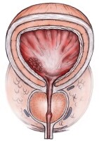
Transurethral Resection of the Prostate

TURP is considered the criterion standard for relieving BOO secondary to BPH. The indications to proceed with a surgical intervention include the following:
  • AUR
  • Failed voiding trials
  • Recurrent gross hematuria
  • Urinary tract infection
  • Renal insufficiency secondary to obstruction
Additional indications for surgical intervention include failure of medical therapy, a desire to terminate medical therapy, and/or financial constraints associated with medical therapy. However, TURP carries a significant risk of morbidity (18%) and mortality risk (0.23%). More recent techniques using bipolar cautery resection devices have lowered the morbidity associated with TURP.
TURP is performed with regional or general anesthesia and involves the placement of a working sheath in the urethra through which a hand-held device with an attached wire loop is placed. High-energy electrical cutting current is run through the loop so that the loop can be used to shave away prostatic tissue. The entire device is usually attached to a video camera to provide vision for the surgeon.
Although TURP is often successful, it has significant drawbacks. When prostatic tissue is cut away, significant bleeding may occur, possibly resulting in termination of the procedure, blood transfusion, and a prolonged hospital stay.
Irrigating fluid may also be absorbed in significant quantities through veins that are cut open, with possible serious sequelae termed transurethral resection syndrome (TUR syndrome). However, this is very rare and does not occur with saline irrigation used in bipolar devices. A urinary catheter must be left in place until the bleeding has mostly cleared.
The large working sheath combined with the use of electrical energy may also result in stricturing of the urethra.
The cutting of the prostate may also result in a partial resection of the urinary sphincteric mechanism, causing the muscle along the bladder outlet to become weak or incompetent. As a result, when the patient ejaculates, this sphincteric mechanism cannot keep the bladder adequately closed. The ejaculate consequently goes backwards into the bladder (ie, retrograde ejaculation), rather than out the penis. Additionally, if the urinary sphincter is damaged, urinary incontinence may result.
The nerves associated with erection run along the outer rim of the prostate, and the high-energy current and/or heat generated by such may damage these nerves, resulting in impotence.
TURP usually requires hospitalization.

Open Prostatectomy

This procedure is now reserved for patients with very large prostates (>75 g), patients with concomitant bladder stones or bladder diverticula, and patients who cannot be positioned for transurethral surgery.
Open prostatectomy requires hospitalization and involves the use of general/regional anesthesia and a lower abdominal incision. The inner core of the prostate (adenoma), which represents the transition zone, is shelled out, thus leaving the peripheral zone behind. This procedure may involve significant blood loss, resulting in transfusion. Open prostatectomy usually has an excellent outcome in terms of improvement of urinary flow and urinary symptoms.
More recently, laparoscopic simple prostatectomy has been performed at a number of institutions and appears to be feasible. However, prostatectomy performed in this fashion still appears to be associated with risk for significant blood loss. Experience to date with this procedure is limited.[27]

Minimally Invasive Treatment

There is considerable interest in the development of other therapies to decrease the amount of obstructing prostate tissue while avoiding the above-mentioned adverse effects associated with TURP. These therapies are collectively called minimally invasive therapies.
Most minimally invasive therapies rely on heat to destroy prostatic tissue. This heat is delivered in a limited and controlled fashion, in the hope of avoiding the complications associated with TURP. They also allow for the use of milder forms of anesthesia, which translates into less anesthetic risk for the patient.
Heat may be delivered in the form of laser energy, microwaves, radiofrequency energy, high-intensity ultrasound waves, and high-voltage electrical energy. As in TURP, delivery devices are usually passed through a working sheath placed in the urethra, although they are usually of a smaller size than that needed for TURP. Devices may also simply be attached or incorporated into a urinary catheter or passed through the rectum, from which the prostate may also be accessed.
Keep in mind that many of these minimally invasive therapies are undergoing constant improvements and refinements, resulting in increased efficacy and safety. Ask urologists about the specifics of the minimally invasive therapies that they use and what results they have experienced.

Transurethral Incision of the Prostate

Transurethral incision of the prostate (TUIP) has been in use for many years and, for a long time, was the only alternative to TURP. It may be performed with local anesthesia and sedation. TUIP is suitable for patients with small prostates and for patients unlikely to tolerate TURP well because of other medical conditions. TUIP is associated with less bleeding and fluid absorption than TURP. It is also associated with a lower incidence of retrograde ejaculation and impotence than TURP.

Lasers

Lasers deliver heat to the prostate in various ways. Lasers heat prostate tissue, causing tissue death by coagulative necrosis, with subsequent tissue contraction; however, laser coagulation of the prostate in this specific sense has met with limited results.
Lasers have also been used to directly evaporate, or to melt away, prostate tissue, which is more effective than laser coagulation. Photoselective vaporization of the prostate produces a beam that does not directly come into contact with the prostate; rather, it delivers heat energy into the prostate, resulting in destruction/ablation of the prostate tissue.
Potassium-titanyl-phosphate (KTP) and holmium lasers are used to cut and/or enucleate the prostate, similar to the TURP technique. These are widely used laser techniques.
Transurethral vaporization/ablation with the KTP or holmium laser can be performed with general or spinal anesthesia and can be performed in an outpatient setting. Catheter time usually lasts less than 24 hours. Studies suggest that photoselective vaporization of the prostate can significantly improve and sustain symptomatic and urodynamic outcomes.
This procedure has been quite useful in patients who require anticoagulation for various medical conditions, since anticoagulation does not need to be interrupted for this procedure, thus further decreasing patient risk.[28, 29]
Lasers may be used in a knifelike fashion to directly cut away prostate tissue (ie, holmium laser enucleation of the prostate), similar to a TURP procedure. The holmium laser allows for simultaneous cutting and coagulation, making it quite useful for prostate resection. Laser enucleation of the prostate has proved to be safe and effective for treatment of symptomatic BPH, regardless of prostate size, with low morbidity and short hospital stay.
TUR syndrome is not seen with this technique, because iso-osmotic saline is used for irrigation. Additionally, removed prostatic tissue is available for histologic evaluation, whereas vaporization/ablation technique does not provide tissue for evaluation. Holmium laser enucleation of the prostate may prove to be the new criterion standard for surgical management of BPH.[29, 30]
Laser treatment usually results in decreased bleeding, fluid absorption, and length of hospital stay, as well as decreased incidence of impotence and retrograde ejaculation when compared with standard TURP. However, healing from laser treatment does not occur until after a period when dead cells slough; thus, patients may experience urinary urgency or irritation, resulting in frequent or uncomfortable urination for a few weeks.
The results of laser therapy vary from one another because not all wavelengths yield the same tissue effects. For example, interstitial lasers (eg, indigo lasers) are designed to heat tissue within the confines of the prostate gland and spread radiant energy at relatively low energy levels. They do not directly involve the urethral portion; thus, irritative symptoms following the procedure are potentially reduced.
Contact lasers such as KTP or holmium, on the other hand, are designed to cut and vaporize at extremely high temperatures They usually bring about more relief of urinary symptoms than treatment with medicines, but not always as much as is provided with TURP. However, KTP laser vaporization and holmium laser enucleation yield results that rival those of TURP.

Transurethral Microwave Therapy

The use of microwave energy, termed transurethral microwave therapy (TUMT), delivers heat to the prostate via a urethral catheter or a transrectal route. The surface closest to the probe (the rectal or urethral surface) is cooled to prevent injury. The heat causes cell death, with subsequent tissue contraction, thereby decreasing prostatic volume.
TUMT can be performed in the outpatient setting with local anesthesia. Microwave treatment appears to be associated with significant prostatic swelling; a considerable number of patients require a urinary catheter until the swelling subsides. In terms of efficacy, TUMT places between medical therapy and TURP. The 2010 AUA guidelines state TUMT is an effective option for partially relieving symptoms that may be considered in patients with moderate or severe LUTS secondary to BPH.[31]

Transurethral Needle Ablation of the Prostate

Transurethral needle ablation of the prostate (TUNA) involves using high-frequency radio waves to produce heat, resulting in a similar process of thermal injury to the prostate as previously described. A specially designed transurethral device with needles is used to deliver the energy.
TUNA can be performed under local anesthesia, allowing the patient to go home the same day. Similar to microwave treatment, radiofrequency treatment is quite popular, and a number of urologists have experience with its use. Radiofrequency treatment appears to reliably result in significant relief of symptoms and better urine flow, although not quite to the extent achieved with TURP. The 2010 AUA guideline update considers TURP an appropriate and effective treatment option for moderate or severe LUTS.[2]

High-Intensity Ultrasound Energy Therapy

High-intensity ultrasound energy therapy delivers heat to prostate tissue, with the subsequent process of thermal injury. High-intensity ultrasound waves may be delivered rectally or extracorporeally and can be used with the patient on intravenous sedation. Urinary retention appears to be common with its use.
High-intensity ultrasound energy also produces moderate results in terms of improvement of the urinary flow rate and urinary symptoms, although its use is now relatively limited compared with the more popular TUNA and TUMT.
High-intensity ultrasound is considered investigational at this time and should not be offered outside of clinical trials.

Mechanical Approaches

Mechanical approaches are used less commonly and are usually reserved for patients who cannot have a formal surgical procedure. Mechanical approaches do not involve the use of energy to treat the prostate.
Prostatic stents are flexible devices that can expand when put in place to improve the flow of urine past the prostate. Their use has been associated with encrustation, pain, incontinence, and overgrowth of tissue through the stent, possibly making their removal quite difficult. To date, their full role and long-term effects are not fully known.

Diet

Data from the Prostate Cancer Prevention Trial revealed that a diet low in fat and red meat and high in protein and vegetables may reduce the risk of symptomatic BPH. Additionally, regular alcohol consumption was associated with a reduced risk of symptomatic BPH, but this is to be interpreted cautiously given the untoward effects of excessive alcohol consumption.[32]

Long-term Monitoring

Patients with BPH who have symptoms significant enough to be placed on medication should be evaluated during office visits to discuss the efficacy of the medication and potential dose adjustment. These visits should take place at least biannually. Patients should undergo DRE and PSA screening at least annually.

Benign Prostatic Hypertrophy Overview

Benign prostatic hyperplasia (BPH), also known as benign prostatic hypertrophy, is a histologic diagnosis characterized by proliferation of the cellular elements of the prostate. Cellular accumulation and gland enlargement may result from epithelial and stromal proliferation, impaired preprogrammed cell death (apoptosis), or both.
BPH involves the stromal and epithelial elements of the prostate arising in the periurethral and transition zones of the gland (see Pathophysiology). The hyperplasia presumably results in enlargement of the prostate that may restrict the flow of urine from the bladder.
BPH is considered a normal part of the aging process in men and is hormonally dependent on testosterone and dihydrotestosterone (DHT) production. An estimated 50% of men demonstrate histopathologic BPH by age 60 years. This number increases to 90% by age 85 years.
The voiding dysfunction that results from prostate gland enlargement and bladder outlet obstruction (BOO) is termed lower urinary tract symptoms (LUTS). It has also been commonly referred to as prostatism, although this term has decreased in popularity. These entities overlap; not all men with BPH have LUTS, and likewise, not all men with LUTS have BPH. Approximately half of men diagnosed with histopathologic BPH demonstrate moderate-to-severe LUTS.
Clinical manifestations of LUTS include urinary frequency, urgency, nocturia (awakening at night to urinate), decreased or intermittent force of stream, or a sensation of incomplete emptying. Complications occur less commonly but may include acute urinary retention (AUR), impaired bladder emptying, the need for corrective surgery, renal failure, recurrent urinary tract infections, bladder stones, or gross hematuria. (See Clinical Presentation.)
Prostate volume may increase over time in men with BPH. In addition, peak urinary flow, voided volume, and symptoms may worsen over time in men with untreated BPH (see Workup). The risk of AUR and the need for corrective surgery increases with age.
Patients who are not bothered by their symptoms and are not experiencing complications of BPH should be managed with a strategy of watchful waiting. Patients with mild LUTS can be treated initially with medical therapy. Transurethral resection of the prostate (TURP) is considered the criterion standard for relieving bladder outlet obstruction (BOO) secondary to BPH. However, there is considerable interest in the development of minimally invasive therapies to accomplish the goal of TURP while avoiding its adverse effects

Anatomy

The prostate is a walnut-sized gland that forms part of the male reproductive system. It is located anterior to the rectum and just distal to the urinary bladder. It is in continuum with the urinary tract and connects directly with the penile urethra. It is therefore a conduit between the bladder and the urethra. (See the image below.)
Normal prostate anatomy. The prostate is located aNormal prostate anatomy. The prostate is located at the apex of the bladder and surrounds the proximal urethra. The gland is composed of several zones or lobes that are enclosed by an outer layer of tissue (capsule). These include the peripheral, central, anterior fibromuscular stroma, and transition zones. BPH originates in the transition zone, which surrounds the urethra.

Pathophysiology

Prostatic enlargement depends on the potent androgen dihydrotestosterone (DHT). In the prostate gland, type II 5-alpha-reductase metabolizes circulating testosterone into DHT, which works locally, not systemically. DHT binds to androgen receptors in the cell nuclei, potentially resulting in BPH.
In vitro studies have shown that large numbers of alpha-1-adrenergic receptors are located in the smooth muscle of the stroma and capsule of the prostate, as well as in the bladder neck. Stimulation of these receptors causes an increase in smooth-muscle tone, which can worsen LUTS. Conversely, blockade of these receptors (see Treatment and Management) can reversibly relax these muscles, with subsequent relief of LUTS.
Microscopically, BPH is characterized as a hyperplastic process. The hyperplasia results in enlargement of the prostate that may restrict the flow of urine from the bladder, resulting in clinical manifestations of BPH. The prostate enlarges with age in a hormonally dependent manner. Notably, castrated males (ie, who are unable to make testosterone) do not develop BPH.
The traditional theory behind BPH is that, as the prostate enlarges, the surrounding capsule prevents it from radially expanding, potentially resulting in urethral compression. However, obstruction-induced bladder dysfunction contributes significantly to LUTS. The bladder wall becomes thickened, trabeculated, and irritable when it is forced to hypertrophy and increase its own contractile force.
This increased sensitivity (detrusor overactivity [DO]), even with small volumes of urine in the bladder, is believed to contribute to urinary frequency and LUTS. The bladder may gradually weaken and lose the ability to empty completely, leading to increased residual urine volume and, possibly, acute or chronic urinary retention.
In the bladder, obstruction leads to smooth-muscle-cell hypertrophy. Biopsy specimens of trabeculated bladders demonstrate evidence of scarce smooth-muscle fibers with an increase in collagen. The collagen fibers limit compliance, leading to higher bladder pressures upon filling. In addition, their presence limits shortening of adjacent smooth muscle cells, leading to impaired emptying and the development of residual urine.
The main function of the prostate gland is to secrete an alkaline fluid that comprises approximately 70% of the seminal volume. The secretions produce lubrication and nutrition for the sperm. The alkaline fluid in the ejaculate results in liquefaction of the seminal plug and helps to neutralize the acidic vaginal environment.
The prostatic urethra is a conduit for semen and prevents retrograde ejaculation (ie, ejaculation resulting in semen being forced backwards into the bladder) by closing off the bladder neck during sexual climax. Ejaculation involves a coordinated contraction of many different components, including the smooth muscles of the seminal vesicles, vasa deferentia, ejaculatory ducts, and the ischiocavernosus and bulbocavernosus muscles.

Epidemiology

BPH is a common problem that affects the quality of life in approximately one third of men older than 50 years. BPH is histologically evident in up to 90% of men by age 85 years. As many as 14 million men in the United States have symptoms of BPH. Worldwide, approximately 30 million men have symptoms related to BPH.
The prevalence of BPH in white and African-American men is similar. However, BPH tends to be more severe and progressive in African-American men, possibly because of the higher testosterone levels, 5-alpha-reductase activity, androgen receptor expression, and growth factor activity in this population. The increased activity leads to an increased rate of prostatic hyperplasia and subsequent enlargement and its sequelae.